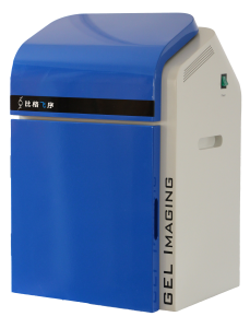
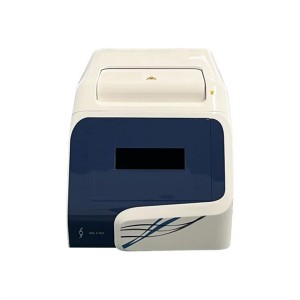
Gel Imaging System
Product features:
Advanced CCD camera
Using the original German imported 16-digit digital CCD camera with high expression and high resolution, low noise and high dynamic range, it can detect DNA/RNA stained with less than 5pg EB, and can identify very close bands and bands with very weak fluorescence intensity.
High transparent digital quantization lens
F/1.2 wide range of zoom capabilities allows for more precise and detailed analysis of specific target areas, providing sharper image quality. Unique lens digital quantization function makes zoom out and aperture size to be adjusted digitally, greatly improve the operating experience, to avoid human error.
The system has the automatic focusing function, avoids the human error.
Camera Obscura
The cabinet panel is formed by the polymer nano-environmental material through the mold once, and the chassis is made of stainless steel once, which ensures the consistency and reliability of the cabinet while ensuring light tightness and anti-interference.
UV SMARTTM no shadow ultra-thin UV transmission table
No light shadow design, brightness and uniformity is much better than the traditional UV transmission table, with a patented gel cutting protection device, protect the body from UV damage.
No damage LED blue/white sample stand
Advanced LED blue light beads, safe and environmentally friendly, no damage to nucleic acid fragments, long-term energy saving and environmental protection.LED white cold light source, toughened glass surface, anti-corrosion and anti-scratch, durable. Magnetic thimble interface, touch control of UV intensity, bring excellent operating experience.
GenoSens image capture software
● Real-time preview of gel images is obtained directly through USB digital interface to facilitate focus
● Advanced pixel merging technology is adopted to improve sensitivity and SNR
● Exposure time or automatic exposure is set by software
● With image rotation, cutting, color inversion and other processing functions to process image optimization
GenoSens image analysis software
● Bands and lanes can be automatically identified, and lanes can be added, removed, and adjusted according to the need to achieve accurate lane separation
● The density integral and peak value of each band in the lane are calculated automatically, which is convenient to calculate the molecular weight and the mobility of each band
● The optical density calculation of the designated area is suitable for quantitative analysis of DNA and protein
● Document management and printing: images in the analysis are saved in BMP format so that the user can terminate or continue the analysis at any time without worrying about the loss of the analysis results. The results of the analysis can be printed by its printing module, including images with analysis identification and user notes, optical density scan images of lane profiles, molecular weight, optical density and mobility analysis results reports
● Analysis result data export: molecular weight, optical density analysis result reports and mobility analysis reports can be exported to text files or Excel files through seamless data linking
Product applications:
Nucleic acid detection:
Fluorescent dyes such as Ethiduim Bromide, SYBRTM Gold, SYBRTM Green, SYBRTM Safe, GelStarTM, Texas Red, Fluorescein, labeled DNA/RNA assay.
Protein detection:
Coomassie bright blue adhesive, silver dyeing adhesive, and fluorescent dyes such as SyproTMRed, SyproTMOrange, Pro-Q Diamond, Deep Purple marker adhesive/membrane/chip etc.
Other applications:
Various hybridization membrane, protein transfer membrane, culture dish colony count, plate, TLC plate.

 中文网站
中文网站
